
Hope Under a Microscope: Check Out UVA’s New $1.25 Million Set of Eyes


George Bloom, far left, wants to better understand Alzheimer’s disease, while Anne Keworthy, far right, will study cholera with the new microscope. Ammasi Periasamy, center, coordinated the grant proposal that led to the microscope’s acquisition. (Photos by Dan Addison, Sanjay Suchak, UVA Health)
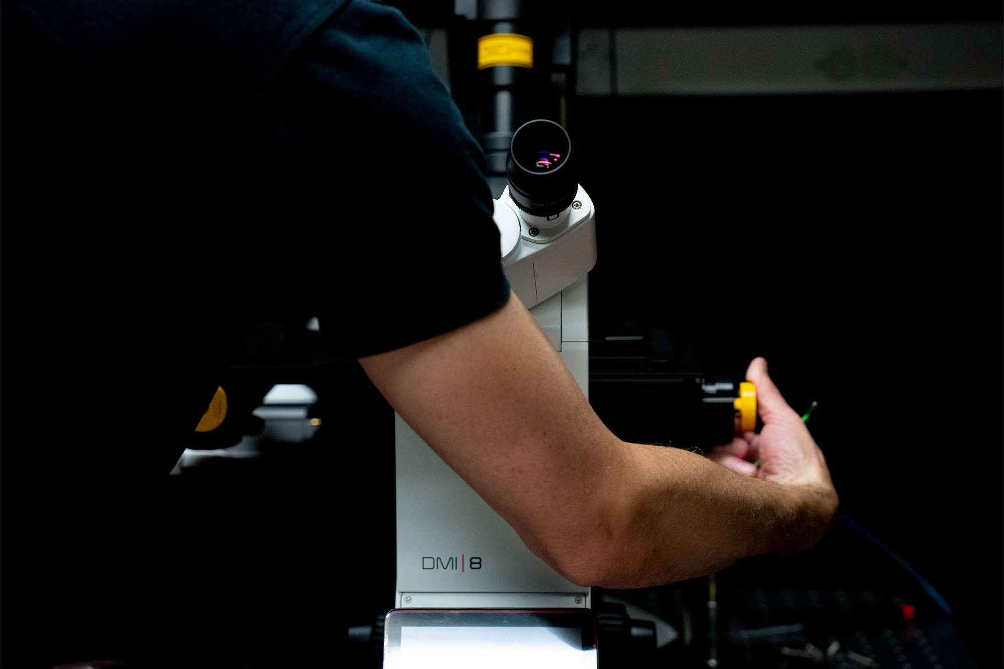
A microscope installer puts the finishing touches on the new Leica microscope. (Photo by Sanjay Suchak, University Communications)
The University of Virginia now has a better set of “eyes” trained on finding answers at the cellular level to problems ranging from Alzheimer’s to infectious bacteria, and many other complex diseases and conditions in between.
With the help of a $1.25 million grant from the National Institutes of Health, UVA acquired a Leica Microsystems Stellaris 8 STED (Stimulated Emission Depletion) super-resolution microscope.
Faculty members say the advanced instrument has the potential to transform their research.
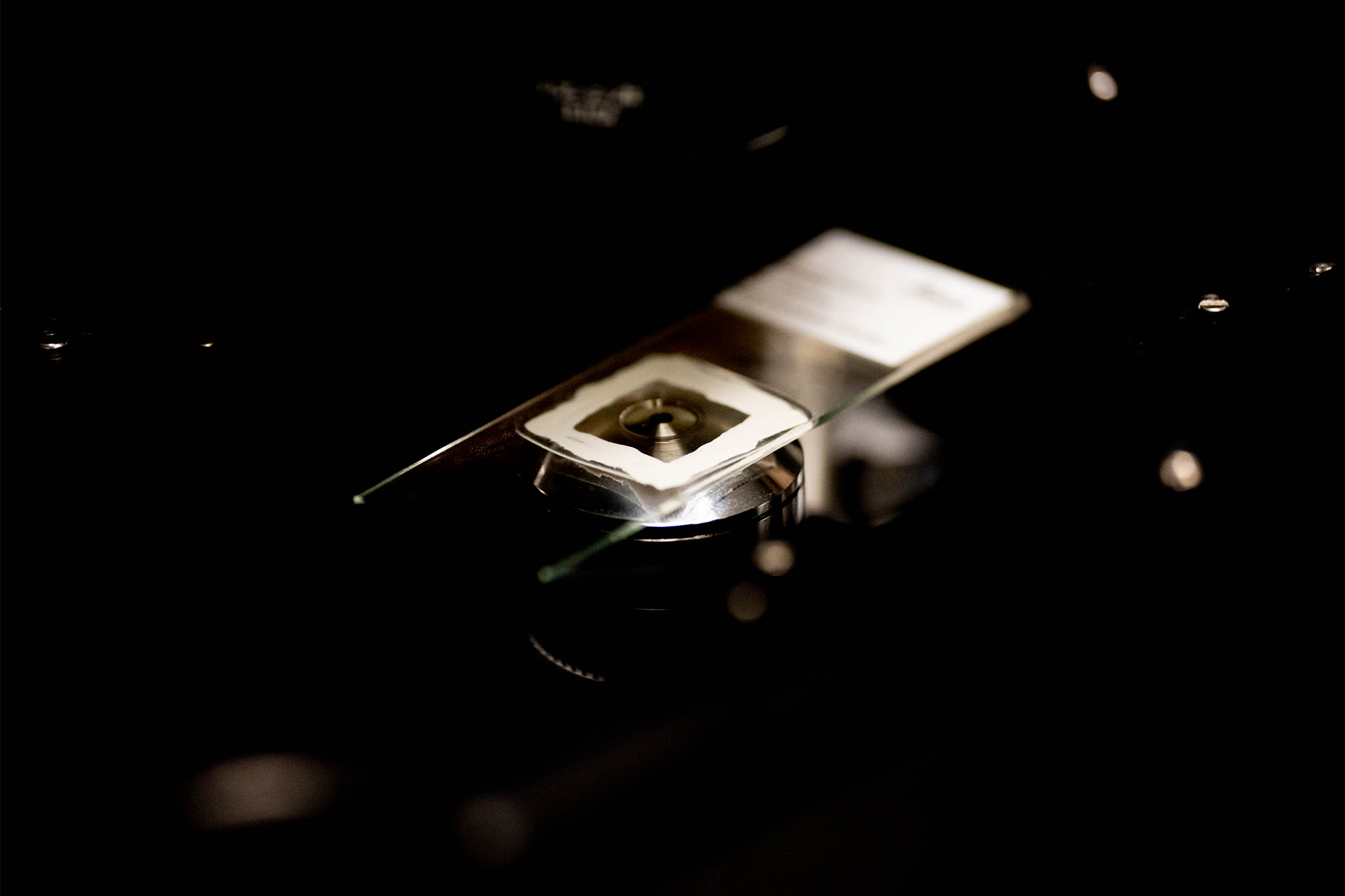
The new microscope uses lasers to deactivate portions of dye-stained biological samples, which minimizes the area of illumination at the focal point and provides a deeper view. (Photo by Sanjay Suchak, University Communications)
Professor George Bloom, an expert in biology, cell biology and neuroscience, said the new tool will facilitate his lab’s research into how subcellular structures within neurons change over the progression of Alzheimer’s disease.
“One of many interesting features of Alzheimer’s disease neurons is that the normally smooth ovoid appearance of their nuclei becomes shriveled and prune-like,” Bloom said. “One of my Ph.D. students, Victoria Sun, discovered what probably causes this phenomenon, and the Leica rep took some beautiful super-resolution images of such a nucleus in a cultured mouse neuron that Victoria treated to become Alzheimer’s disease-like.”
The new system, the latest addition to the W.M. Keck Center for Cellular Imaging, was installed at the end of September and now allows users to see down to 20- to 30-nanometer resolution. To put that into perspective, a sheet of paper is 100,000 nanometers thick.
What’s more, the microscope can produce both two- and three-dimensional images of live cells at between 30- and 70-nanometer resolution.
That might mean the difference in a researcher knowing if a protein was even expressed in a biological sample, said Keck Center director Ammasi Periasamy, a professor of biology and biomedical engineering.
“Now they see it; before, they couldn’t,” said Periasamy, who applied for the NIH Office of the Director grant in association with about a dozen UVA researchers who will be using the microscope.
Among those professors is Anne Kenworthy, the assistant director of the Center for Membrane and Cell Physiology, Molecular Physiology and Biological Physics. Her group is studying nanoscale complexes of proteins and lipids that cluster together in cell membranes known as rafts.
“We are trying to understand how cholera toxin, the bacterial toxin that causes cholera, binds to and manipulates rafts to force its way into cells,” Kenworthy said.
Using the microscope to gain a better understanding of what’s happening at the cellular level, she hopes to identify pharmacological tools to either stabilize or disrupt the rafts, depending on the body’s needs at a given time.
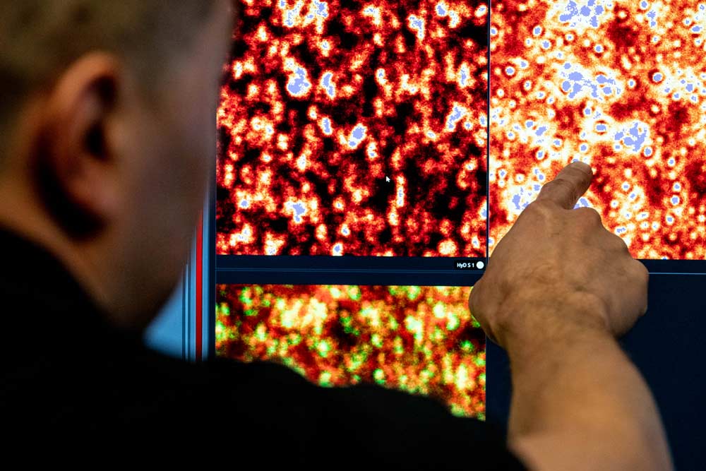
A microscope installer demonstrates the high-resolution capability of the new system. (Photo by Sanjay Suchak, University Communications)
To facilitate their viewing, scientists are able to stain cell components with fluorescent markers. The new microscope works by allowing the machine to both “see” the marker as if being viewed by a standard, high-powered confocal microscope, but also to look deeper.
To get beyond the limitations of standard light diffraction, the microscope uses lasers to deactivate part of the dye labeling, thereby minimizing the area of illumination at the focal point. That enhances resolution for the remaining labeled proteins.
The College and Graduate School of Arts & Sciences provided $150,000 in matching funds for the new system.
UVA graduate and undergraduate students will also be able to use the machine once they’ve gone through the appropriate training. For information, contact Periasamy via the Keck Center.


We’re here to answer your questions! Contact us today.